

Main content 1 Menu 2 Search 3 Footer 4
+A
A
-A
High contrast
HOME JOURNAL JOURNAL SELECTION NETWORK HELP ABOUT

Journal Selection Criteria and Standards

WPRIM Journal Selection Criteria (August 2023)

NJSC Philippines Selection Criteria (for Philippine-based journals only)

Minimum standards for the suspension and removal of WPRIM approved journals

Application and Indexing Process

Application and Submission Process for WPRIM Indexing

Journal Content Management

Candidate Journal Selection and Data Creation and Management System

Journal of Veterinary Clinics

2001  (1,  18)  to  Present  ISSN: 1598-298X

Articles

About

Save Email

Sort by

Best match
Relevance
PubYear
JournalTitle

DISPLAY OPTIONS

Format:

Per page:

Save citations to file

Selection:

Format:

Create file Cancel

Email citations

To:

Please check your email address first!

Selection:

Format:

Send email Cancel

93

results

page

of 10

1

Cite

Cite

Copy

Share

Share

Copy

Computed Tomography and Magnetic Resonance Imaging Features of Spinal Chondrosarcoma in a Cat

Minhee LEE ; Sang-Kwon LEE ; Juyoung SHIN ; Seulgi BAE ; Kija LEE

Journal of Veterinary Clinics.2024;41(2):133-138. doi:10.17555/jvc.2024.41.2.133

An 8-year-old, spayed female Persian cat weighing 3.6 kg presented with a lumbosacral mass and bilateral weight bearing hindlimb lameness. Computed tomography (CT) and magnetic resonance imaging (MRI) revealed a dumbbell-shaped heterogeneous mass extending through the internal surface of the ileum and surrounding the lumbosacral junction. CT also revealed extensive osteoproliferation and bone lysis of the sacrum, but no evidence of any pulmonary metastasis. Furthermore, MRI revealed a focal area in the spinal cord showing connection with the adjacent tumor, suggesting tumor invasion into the spinal cord. Low-grade myxoid chondrosarcoma was histopathologically diagnosed. This is the first report describing CT and MRI findings of spinal cord chondrosarcoma in veterinary medicine. This study suggests that combining CT with MRI is a more sensitive tool for evaluating spinal tumors than using CT or MRI alone.
2

Cite

Cite

Copy

Share

Share

Copy

Asymptomatic Uterine Rupture Caused by Trauma in a Small-Sized Non-Gravid Dog

Ye-Eun KIM ; Sang-Hyun NAM ; Won-Jong LEE ; Chang-Hwan MOON ; Geum-Lan HONG ; Ju-Young JUNG ; Jae-Min JEONG ; Hae-Beom LEE ; Seong-Mok JEONG ; Dae-Hyun KIM

Journal of Veterinary Clinics.2024;41(2):112-116. doi:10.17555/jvc.2024.41.2.112

Uterine rupture is uncommon in small animals and can be caused by trauma, infection, dead fetus, or inappropriate obstetric techniques. A 4-yearold, intact female mixed-breed dog weighing 3 kg presented for elective ovariohysterectomy with a history of a motor vehicle accident two months previously. The patient showed no clinical signs of uterine abnormalities or evidence of pregnancy during physical examination. Clinical examinations, radiography, and ultrasonography revealed no significant findings. During ovariohysterectomy, a completely transected left uterine horn and dome-shaped ends were identified.The uterine layers were everted such that the endometrium could be seen outside the uterine horn. The dog recovered without post-operative complications and was discharged the following day. This report describes a rare case of an asymptomatic uterine rupture caused by trauma in a non-gravid dog.

3

Cite

Cite

Copy

Share

Share

Copy

Successful Management of Post-Attenuation Neurologic Signs and Portal Hypertension in a Dog with Congenital Portoazygos Shunt

Jin-Young KIM ; Kun-Ho SONG ; Joong-Hyun SONG

Journal of Veterinary Clinics.2024;41(2):95-100. doi:10.17555/jvc.2024.41.2.95

A 6-year-old neutered male Poodle with a body weight of 2.7 kg was diagnosed with a congenital extrahepatic portoazygos shunt presented with progressive hepatic encephalopathy. Five days after surgical attenuation, the patient showed post-attenuation neurological signs (PANS) such as generalized tonic-clonic seizure, dull mentation, and tremor. PANS were successfully managed with antiepileptic drugs, but third-space fluid accumulation (ascites and peripheral edema) and phlebectasia were newly identified, suggesting marked portal hypertension (PHT). Telmisartan, spironolactone, carvedilol, and prednisolone were sequentially administered. Three months after surgery, both abnormal clinicopathological values such as anemia and hypoalbuminemia, and clinical signs completely resolved. Herein, we report successful management strategies for PANS and PHT in a dog following surgical attenuation of a congenital portoazygos shunt.

4

Cite

Cite

Copy

Share

Share

Copy

Large Mesenteric Pedunculated Lipoma in a Horse

Seokho SON ; Seyoung LEE ; Eun-bee LEE ; Kyung-won PARK ; Ji-Youl JUNG ; Jae-Hoon KIM ; Hyohoon JEONG ; Jong-pil SEO

Journal of Veterinary Clinics.2024;41(2):123-126. doi:10.17555/jvc.2024.41.2.123

A seven-month pregnant 15-year-old Thoroughbred mare presented with acute abdominal pain to Jeju National University Equine Hospital. At presentation, a nasogastric intubation revealed 10 L of gastric reflux; rectal palpation and ultrasound revealed dilated loops and thickening of the walls of the small intestine. An exploratory laparotomy revealed strangulation of the small intestine due to a large abdominal mass. The mass was double-ligated and resected blindly due to the short pedicle. An enterectomy was not performed as intestinal motility was detected following the mass removal. Histopathological examination confirmed that the mass was a lipoma, measuring 24 cm × 16 cm × 16 cm in size. On day 8 post-surgery, the mare was discharged without complications. This case report describes the diagnosis and treatment of strangulation of the small intestine by a pedunculated lipoma, thus providing useful information on lipoma in horses.

5

Cite

Cite

Copy

Share

Share

Copy

Magnetic Resonance Imaging and Ultrasonographic Evaluation of Canine Tarsus

Soomin PARK ; Sang-hwa RYU ; Jae-gwan HEO ; Eun-jee KIM ; Jihye CHOI ; Junghee YOON

Journal of Veterinary Clinics.2024;41(2):79-87. doi:10.17555/jvc.2024.41.2.79

The tarsus in dogs has a complex structure that makes its evaluation relatively challenging. Because an accurate diagnosis of the tarsus is difficult through basic examinations alone, imaging tests are essential. Previous studies have explored the anatomical and radiological features of the canine tarsus using several imaging modalities. However, the imaging utility of the tarsus across different modalities has not been thoroughly evaluated. This study aimed to visualize the tarsal structures using magnetic resonance imaging (MRI) and ultrasonography, compare their utility, and propose suitable imaging modalities and conditions for evaluating specific tarsal structures. Magnetic resonance imaging and ultrasound scans of the tarsus of four healthy dogs were performed, and two observers rated the utility of each image on a five-point scale. Although MRI is more beneficial for assessing the tarsal structures than ultrasound, ultrasound also appears clinically useful for evaluating the cranial tibialis muscle, deep digital flexor tendon, subcutaneous fat, joint space, and superficial digital flexor tendon. In addition, each structure of interest can be evaluated for optimal visibility using specific ultrasound sections, MRI sequences, and planes. In veterinary clinical practice, an initial assessment using ultrasound imaging with optimal visibility is required and if further evaluation is necessary, MRI examinations with optimal MRI sequences and planes can be performed.

6

Cite

Cite

Copy

Share

Share

Copy

Transient Myocardial Thickening in a 4-year-old Korean Domestic Shorthair Cat

Yunhee JOUNG ; Hyerin AHN ; Jeongbae CHOI ; YoungMin YUN ; Woo-Jin SONG

Journal of Veterinary Clinics.2024;41(2):106-111. doi:10.17555/jvc.2024.41.2.106

A 4-year-old neutered female domestic shorthair cat weighing 5.1 kg was referred to Jeju National University Hospital with acute onset respiratory distress, weakness, and anorexia. The patient had a history of stressful antecedent events that involved bullying by a newly introduced cat. Thoracic radiography and echocardiography revealed a stage C hypertrophic cardiomyopathy phenotype based on the American College of Veterinary Internal Medicine classification system with pulmonary edema, pleural effusion, and pericardial effusion at the same time. The patient was treated with furosemide, pimobendan, and rivaroxaban. Pericardiocentesis was performed because pericardial effusion was identified. Reevaluation after 30 days revealed a normal respiratory rate on physical examination, normal cardiac shape on thoracic radiographs, and normal cardiac measurements on echocardiography. The patient was tentatively diagnosed with transient myocardial thickening (TMT) and all medications were discontinued. Six months after the initial hospitalization, the cat continued to do well without any clinical signs or left ventricular wall thickening. This case is the first report describing feline TMT in Korea. Moreover, it involves a rare case in which pulmonary edema, pleural effusion, and pericardial effusion, which induce cardiac tamponade, occurred simultaneously due to TMT-related congestive heart failure.

7

Cite

Cite

Copy

Share

Share

Copy

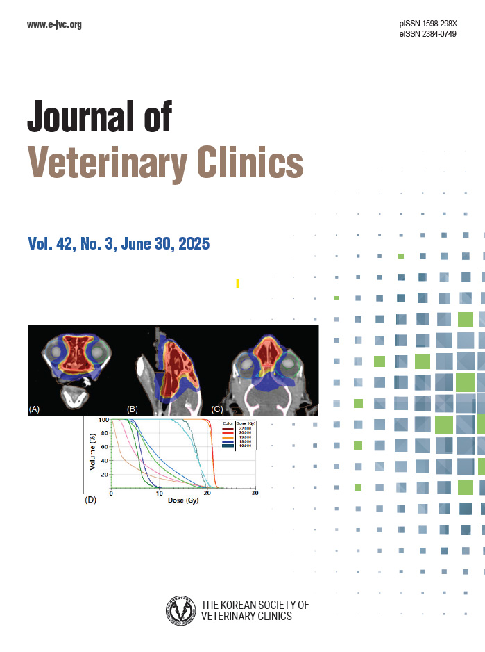
Successful Treatment of Feline Nasopharyngeal Lymphoma by Hypofractionated Radiation Therapy After Surgical Debulking in a Cat

Sumin KIM ; Gunha HWANG ; Jin-Yoo KIM ; Chi-Oh YUN ; Seunghwa LEE ; Moonyeong CHOI ; Joong-Hyun SONG ; Hee Chun LEE ; Tae Sung HWANG

Journal of Veterinary Clinics.2024;41(2):117-122. doi:10.17555/jvc.2024.41.2.117

A 3-year-old spayed female Russian blue cat was presented for dyspnea, nasal discharge, and stertorous breathing. Plain thoracic radiography revealed no specific findings. Computed tomography (CT) was performed to differentiate upper airway tract disorders. It revealed the presence of an iso-attenuating mass measuring 10.0 × 7.9 × 15.6 mm, with mild homogeneous contrast enhancement occupying the rostral nasopharynx. The mass was surgically debulked via a longitudinal incision in the soft palate. Histopathological and immunohistochemistry analysis of the surgically excised mass revealed CD3–/ CD79a+ B cell lymphoma with an incomplete margin. The patient underwent hypofractionated radiation therapy, receiving a total of 36 Gray (Gy) in 6 Gy fractions over a six-week period. A follow-up CT examination was performed after 27 months of irradiation and the patient was confirmed to have achieved a complete response. There were no complications related to irradiation. The patient was alive for 40 months without recurrence. This study suggests that hypofractionated radiation therapy combined with surgical debulking could be considered as a treatment option for feline nasopharyngeal lymphoma.

8

Cite

Cite

Copy

Share

Share

Copy

Diagnosis of Coxofemoral Joint Luxation in a Whooper Swan (Cygnus Cygnus) Using Computed Tomography and Radiography

Jinho JANG ; Jong-pil SEO ; Hyohoon JEONG ; Seyoung LEE ; YoungMin YUN

Journal of Veterinary Clinics.2024;41(2):139-142. doi:10.17555/jvc.2024.41.2.139

A wild Whooper swan (Cygnus Cygnus) with limping due to an injured left pelvic limb in an accident was rescued on the seashore and transferred to the Jeju Wildlife Rescue Center on November 23rd, 2020. On physical examination, its body condition score was 1 out of 5 due to starvation and dehydration.The left coxofemoral joint was also examined by careful palpating and estimating the damage. Moderated soft tissue swelling and crepitus surrounding the hip joint were confirmed. Radiography and computed tomography (CT) were used together for an accurate diagnosis of the joint. By radiographs readings, it was difficult to accurately confirm the condition of the proximal femur due to superimposition of the synsacrum and internal organs. However, signs such as avulsion fracture of the femoral head and a few fragments around the joint were revealed by CT imaging. Besides, through three-dimensional (3D) image analysis of CT, the dislocated area and condition of the left hip joint could be accurately and easily confirmed. The diagnostic process showing in this paper could be used as a good reference for diagnosing coxofemoral joint luxation in wild swan.

9

Cite

Cite

Copy

Share

Share

Copy

Morphologic and Molecular Characterization of Psoroptes ovis from Pet Rabbits in South Korea

Md Ashraful ISLAM ; Obaidul ISLAM ; Md Sodrul ISLAM ; Sungryong KIM ; Mohammed MEBAREK BIA ; Seongjun CHOE ; Ki–Jeong NA

Journal of Veterinary Clinics.2024;41(2):88-94. doi:10.17555/jvc.2024.41.2.88

Pet rabbits are affected by the highly contagious ectoparasite Psoroptes (P.) ovis, which carries significant economic implications for the global rabbit industry. Accurate identification of the mite species remains essential to implement effective treatment and control strategies. Two approximately oneyear-old female pet rabbits were admitted to the Veterinary Teaching Hospital of Chungbuk National University due to excessive scratching of the ears and the presence of waxy debris within the ear canals. Mites were isolated from the waxy debris extracted from the ear canals and subsequently identified as Psoroptes spp. through microscopic examination. Species confirmation was achieved through mitochondrial cytochrome c oxidase subunit 1 cox1 gene analysis. The analysis revealed the mites to be P. ovis based on cox1 gene sequences. The deposited GenBank accession numbers for these sequences are OR985022 and OR985023. This represents the first report of mitochondrial cox1 gene sequences of P. ovis isolated from pet rabbits in South Korea.

10

Cite

Cite

Copy

Share

Share

Copy

Bowel Obstruction Due to Less-Digestible Pet Food with No Shadowing on Ultrasonography in a SmallBreed Puppy

Junyoung KIM ; Jihye CHOI ; Junghee YOON

Journal of Veterinary Clinics.2024;41(2):127-132. doi:10.17555/jvc.2024.41.2.127

A 6-month-old intact female Bichon Frise dog weighing 0.9 kg presented with vomiting, anorexia, and lethargy persisting for 3 days. No remarkable abnormalities were detected on the history or physical examination. Laboratory findings were mostly normal, except for elevated levels of alkaline phosphatase (ALP) and blood urea nitrogen (BUN). Abdominal radiography revealed a fluid-dilated stomach and gas-dilated intestinal loops in the regional areas. Abdominal ultrasonography was performed to investigate the cause of gastrointestinal dilation, which revealed a rectangular, homogeneous, echogenic foreign material with no shadowing in the small intestine, causing mechanical obstruction. Upon further inquiry involving detailed re-take of history with the owner, a history of ingesting dog gum 4 days prior was identified. On surgical enterotomy, the hard pet food was identified and removed from the distal duodenum. Postoperatively, the patient’s clinical signs showed complete improvement, with a return to normal appetite. The present case demonstrates that less-digestible, hard pet food, despite showing no shadowing on ultrasonography, can act as a foreign material, causing mechanical intestinal obstruction in a small-breed puppy. Furthermore, surgical removal of these materials is necessary in cases of intestinal obstruction.

Country

Republic of Korea

Publisher

The Korean Society of Veterinary Clinics

ElectronicLinks

https://www.e-jvc.org/main.html

Editor-in-chief

Ki-Jeong Na

E-mail

editor@e-jvc.org

Abbreviation

J Vet Clin

Vernacular Journal Title

ISSN

1598-298X

EISSN

2384-0749

Year Approved

2025

Current Indexing Status

Currently Indexed

Start Year

2001

Description

Journal of Veterinary Clinics (J Vet Clin, JVC) is an open access, peer-reviewed, online-only veterinary medicine journal published in English bimonthly (Febraury 28, April 30, June 30, August 31, October 31, and December 31: 6 times per year), which is an official journal of the Korean Society of Veterinary Clinics. The journal was first published as name Korean Journal of Veterinary Clinical Medicine (ISSN 1225-4800) in 1984 and has been changed the name as Journal of Veterinary Clinics (ISSN 1598-298X) since 2001. JVC is an international journal which publishes papers on all aspects of the veterinary clinics. Clinical case reports, scientific research papers, short communications, notes, and review articles covering the diagnosis, treatment, and control of animal disease will be considered. In addition to members of the JVC, any researchers throughout the world can submit a manuscript if the scope of the manuscript is appropriate. JVC makes it a priority to keep the entire process of submission, review, and publication as fast as possible while maintaining the best of international standard rigor. Therefore, JVC offers online manuscript submission for rapid peer-review and communication with authors. This journal is supported by the Korean Federation of Science and Technology Societies (KOFST) Grant funded by the Korean Government. All or part of the content is indexed/tracked/covered by SCOPUS, KCI, KSCI, KISTI, CrossRef, Google Scholar, Naver Academic.

Related Sites

WHO WPRO GIM

Help Accessibility
DCMS Web Policy
CJSS Privacy Policy

Powered by IMICAMS( 备案号: 11010502037788, 京ICP备10218182号-8)

Successfully copied to clipboard.