

Main content 1 Menu 2 Search 3 Footer 4
+A
A
-A
High contrast
HOME JOURNAL JOURNAL SELECTION NETWORK HELP ABOUT

Journal Selection Criteria and Standards

WPRIM Journal Selection Criteria (August 2026)

NJSC Philippines Selection Criteria (for Philippine-based journals only)

Minimum standards for the suspension and removal of WPRIM approved journals

Application and Indexing Process

Application and Submission Process for WPRIM Indexing

Journal Content Management

Candidate Journal Selection and Data Creation and Management System

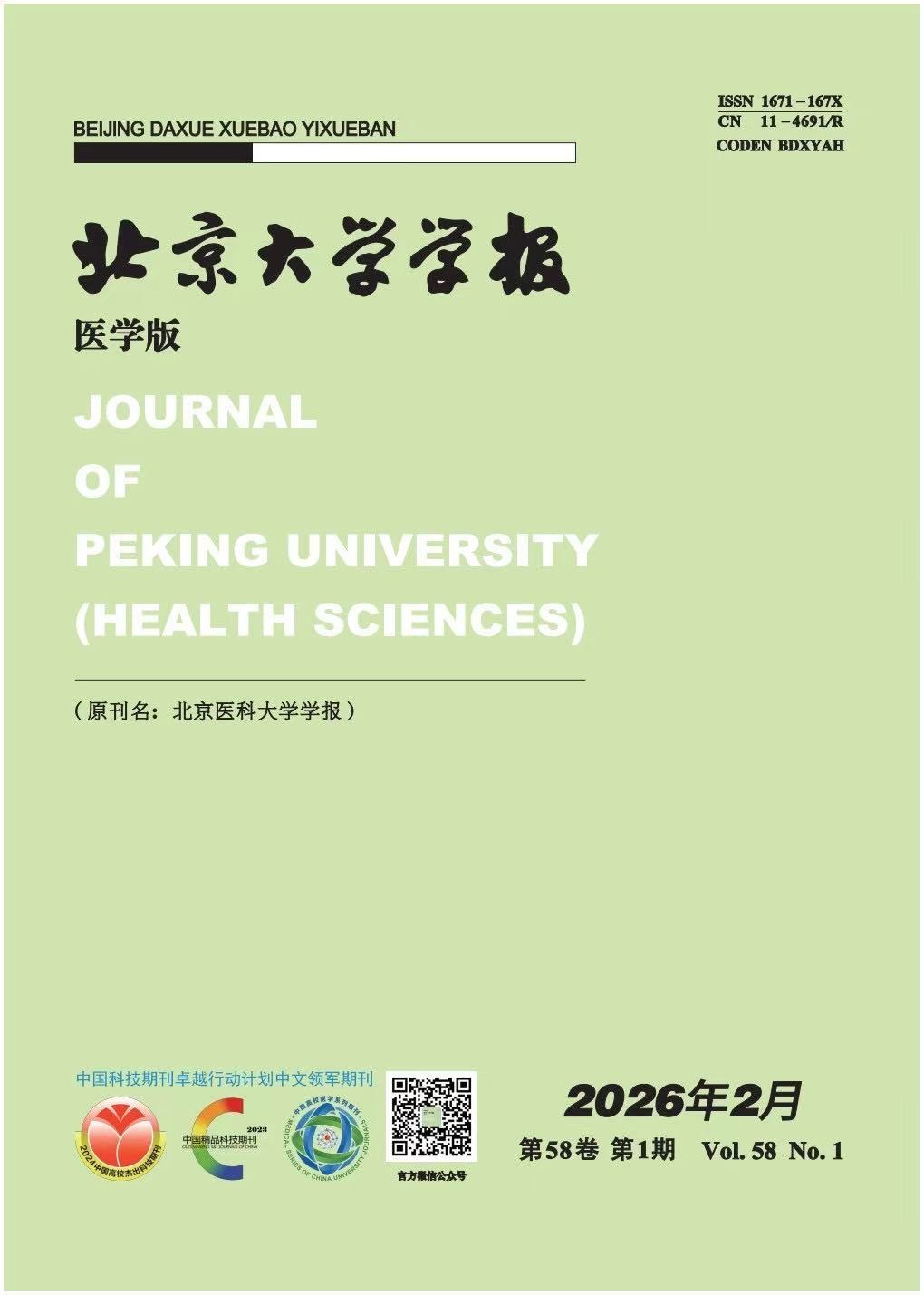
Journal of Peking University(Health Sciences)

1959  to  Present  ISSN: 1671-167X

Articles

About

Save Email

Sort by

Best match
Relevance
PubYear
JournalTitle

DISPLAY OPTIONS

Format:

Per page:

Save citations to file

Selection:

Format:

Create file Cancel

Email citations

To:

Please check your email address first!

Selection:

Format:

Send email Cancel

2645

results

page

of 265

1

Cite

Cite

Copy

Share

Share

Copy

Distant metastases of malignant salivary gland carcinoma after treated by 125Ⅰ internal brachy therapy alone

Wei WANG ; Lei ZHENG ; Shuming LIU ; Mingwei HUANG ; Yan SHI ; Xiaoming LV ; Jie ZHANG ; Jianguo ZHANG

Journal of Peking University(Health Sciences).2017;49(3):547-550. doi:10.3969/j.issn.1671-167X.2017.03.029

Objective: To investigate clinical features of distant metastases from malignant salivary gland carcinomas after treated by 125I internal brachy therapy alone.Methods: Between 2002 and 2010, 43 patients with locally advanced unresectable or recurrent malignant salivary gland carcinomas were treated by 125I internal brachy therapy alone at Peking University School and Hospital of Stomatology.All of them had been follow-up at least 2 years.The primary sites of malignant salivary gland carcinomas were the parotid for 12 patients, infratemporal fossa for 9 patients, tongue for 7 patients, maxilla for 6 patients, parapharyngeal for 4 patients, floor of months for 3 patients, submandibular gland for 1 patient, and lip for 1 patient.The overall survival rate, local control rate, and distant metastases were retrospectively reviewed.Results: Distant metastases occurred in 23 of the 43 patients (53.5%).Distant metastases developed from 5 to 96 months, with an average interval of (27.0±23.7) months from the time of initial diagnosis, the mean interval was 21 months.The commonest site of distant metastases overall was the lung 69.6%(16/23).The most common pathological types of distant metastases were adenoid cystic carcinoma (14/23, 60.9%) and nonspecific adenocarcinoma (7/23, 30.4%).At the time of the last follow-up, 26 patients died, and 18 of them due to distant metastases (69.2%, 18/26).In the study, 27 patients got complete remission(CR), 12 got partial remission(PR) more than 50%, 2 less than 50%, and 2 patients were invalid.The effective rate (CR+ PR) was 90.7%.The 3 year loco-regional control rate and survival rate were 60.1%, 82.6%, respectively, and the 5 year's 53.4% and 56.0%.Conclusion: The 125I brachy therapy is a feasible and effective modality for the treatment of locally advanced unresectable or recurrent malignant salivary gland carcinomas.However, further studies are needed with larger numbers of patients and for a longer follow-up assessment.Distant metastasis was the main cause of treatment failure, and the lung was the most common site of distant metastases.
2

Cite

Cite

Copy

Share

Share

Copy

Detection and management of the vascular paths in the lateral sinus wall using cone beam computed tomography

Quan CHEN ; Xiao ZHANG ; Zhiyong ZHANG ; Wei GAO ; Wenshu LIU ; Tian MENG ; Yuhuan CHEN ; Huili WANG

Journal of Peking University(Health Sciences).2017;49(3):540-546. doi:10.3969/j.issn.1671-167X.2017.03.028

Objective: To detect the vascular paths in the lateral wall of maxillary sinus using cone beam computer tomography (CBCT), and to retrospect the surgical managements of avoiding bleeding complication during the lateral approach maxillary sinus elevation.Methods: The documents of 71 consecutive patients with 81 sides maxillary sinus elevation surgery were collected.The vascular paths in the lateral wall of maxillary sinus were detected by the preoperative CBCT, and the messages about the vascular in surgical records were analyzed.Results: The paths of the vascular could be detected in 77 (95.1%) sides maxillary sinus in the reconstruction panoramic images of CBCT.At the position of the first molar, the paths of the vascular of the lateral maxillary sinus walls could be detected in 54 sides (66.7%) in the reconstruction coronal images of CBCT, and the other 27 sides (33.3%) could not be detected.Two approximately parallel paths of the vascular were found in 3 sides (3.7%) of the lateral maxillary sinus walls.The different diagnoses occurred in 6 sinuses between two observers.The kappa of diagnostic consistency of the two observers was 0.842 (P<0.001).The mean distance between the lower border of the vascular path to the plane of the alveolar crest of 54 sides maxillary sinuses was about (13.0±4.7) mm.The mean distance between lower border of vascular path to the plane of the floor of the sinus was (9.3±4.8) mm.The vascular path was located in the floor wall in 1 sinus.During the lateral approach maxillary sinus elevation operation, intraosseous vessels were dissected in 4 sides sinus lateral wall, the vascular path was avoided consciously in 3 sides, and the sinus elevation surgery had to be given up in 1 side for the vessel was torn and bleeding.There were no vascular related messages in 73 sides of the lateral approach maxillary sinus elevation operation records.Conclusion: The vascular paths of maxillary sinus wall could be detected by CBCT in most cases.Preoperative CBCT examination was proved to be reliable.The vascular paths of maxillary sinus wall should be examined carefully.It was helpful to make the surgical design perfectible and reduce the risk of tearing the vessel in operation.

3

Cite

Cite

Copy

Share

Share

Copy

Diagnosis accuracy of fine-needle aspiration cytology for vascular anomalies

Yunan LIU ; Xiaojing LIU ; Xiaomeng YU ; Fuyun ZHAO

Journal of Peking University(Health Sciences).2017;49(3):527-530. doi:10.3969/j.issn.1671-167X.2017.03.026

Objective:To evaluate the feasibility of fine-needle aspiration cytology (FNAC) in diagnosing vascular abnormality in oral and maxillofacial region.Methods: The method of retrospective study was used.The data from the patients who underwent FNAC from 2011 to 2014 in Department of Oral and Maxillofacial Surgery, Peking University School of Stomatology were collected.All the included patients were divided into surgery group and non-surgery group.The patients in surgery group underwent lesion resection and the postoperative pathological results were gained.The patients in non-surgery group underwent periodical sclerotherapy.The accuracy of FNAC was identified by histopathologic diagnosis in surgery group and the effect of sclerotherapy in non-surgery group.Results: In this study, 93 patients were involved, including 51 males and 42 females.The median age was 2.5 years.Among them, 67 cases were judged as vascular abnormality by FNAC, and 63 cases were in consistent with final diagnosis and 4 cases were not.Among the other 26 cases which were diagnosed as other diseases by FNAC, 5 cases were accorded with final diagnosis.Therefore, the sensitivity of FNAC on diagnosis of vascular abnormity was 93% and its specificity was 84%.Among them, there were 29 cases in the surgery group and 64 cases in the non-surgery group.FNAC results were in consistent with the postoperative pathological results in 20 cases in surgery group (69%), and not consistent with pathological results in 9 cases.The pathological diagnoses included vascular malformations (3 cases), neurofibromas (2 cases), hamartoma (1 case), sebaceous cyst (1 case), adenolymphoma (1 case), and descriptive diagnosis (1 case).In non-surgery group, FNAC results were in consistent with the clinical effect of sclerotherapy in 61 cases (95%).There were 3 misdiagnosed cases.The coincident rate between the result of FNAC and that of pathological or clinical diagnosis was 86%.Conclusion: FNAC is a feasible and minimal invasive method to diagnose vascular abnormality in oral and maxillofacial region.

4

Cite

Cite

Copy

Share

Share

Copy

Effects of PTCH1 mutations on the epithelial proliferation derived from keratocystic odontogenic tumour

Zihan SIMA ; Yingying HONG ; Tiejun LI

Journal of Peking University(Health Sciences).2017;49(3):522-526. doi:10.3969/j.issn.1671-167X.2017.03.025

Objective: To explore the relationship between the PTCH1 mutation and the expression of bcl-2, filaggrin, and loricrin in the keratocystic odontogenic tumour (KCOT), as well as the effects of the mutated PTCH1 on the epithelial proliferation and differentiation.Methods: The samples were collected from 20 cases of KCOT with mutated PTCH1, as well as 20 cases without mutation.All the samples were analyzed with immunohistochemical staining, for the purpose of investigating the expression of bcl-2, filaggrin, and loricrin.Results: In the samples with mutated PTCH1, the epithelia of 60% (12/20) cases expressed intensively positive bcl-2 staining, 20% (4/20) expressed moderate staining, and 20% (4/20) weak staining, but no negative bcl-2 staining samples were investigated;it was significantly different from the samples without PTCH1 mutation, in which 20% (4/20) expressed intensive staining, no moderate staining, 50% (10/20) weak staining, and 30% (6/20) negative staining were investigated (U=72, P=0.001).For the expression of filaggrin, 55% (11/20) of samples with PTCH1 mutations were stained weakly and 45% (9/20) showed negative staining, while in the samples not harboring PTCH1 mutations, 30% (6/20) cases showed moderate positive staining, 40% (8/20) weak staining and 30% (6/20) negative staining, no intensive staining was investigated (U=182, P=0.48).The loricrin expressed in all the layer of the epithelia in all the samples, while the filaggrin was mainly loca-lized within 1-4 layer cells of the epithelia.The differences of the expression of filaggrin and loricrin between the samples with mutated PTCH1 and without mutated PTCH1 displayed no significance.Conclusion: In the epithelia of KCOT, the bcl-2 expression was significantly associated with the PTCH1 mutation, which suggested that the mutated PTCH1 gene perhaps promotes the proliferation of KCOT epithelium.

5

Cite

Cite

Copy

Share

Share

Copy

Biomechanical analysis of the deep radioulnar ligaments stabilizing the distal radioulnar joint

Yanbo RONG ; Guanglei TIAN ; Shanlin CHEN

Journal of Peking University(Health Sciences).2017;49(3):518-521. doi:10.3969/j.issn.1671-167X.2017.03.024

Objective: To evaluate the role of the deep radioulnar ligament in the stability of the distal radioulnar joint (DRUJ).Methods: In the study, 14 fresh cadaver upper extremities were randomly divided into two groups.After exposuring the palmar and dorsal deep distal radioulnar ligament, one group was marked as palmar deep radioulnar ligament, and the other group was marked as dorsal deep radioulnar ligament.The pronator teres and the supinator were exposed.A Kirschner wire perpendicular to the bone on Lister tubercle of radius was inserted, then another Kirschner wire on the same level of ulnar inserted when the forearm was in neural position, which was kept parallel to the first Kirschner wire.These specimens were mounted on a specially designed jig which held the limb rigidly, keeping the elbow fle-xion and the ulnar fixation.The radius could freely rotate around the ulnar.Then 50 N force on the pronator teres was applied to simulate the active pronation, and 60 N force on the supinator to simulate the active supination.The active pronation was stimulated, and the displacement of the distal radius was measured with respect to the ulna.The active supination was atimulated, and the displacement of the distal radius was measured with respect to the ulna.The palmar deep radioulnar ligament in one group was cut, then the displacement of the distal radius measured with respect to the ulna when the forearm was in pronation and supination.The dorsal deep radioulnar ligament in the other group was cut, and the displacement of the distal radius measured with respect to the ulna when the forearm was in pronation and supination.Results: After resection of the palmar deep radioulnar ligament, the displacement of the distal radius with respect to the ulna was statistically significantly different when the forearm was in pronation (t=5.591, P=0.001), but there was no difference when the forearm was in supination (t=0.433, P=0.680).After resection of the dorsal deep radioulnar ligament, the displacement of the distal radius with respect to the ulna was not different when the forearm was in pronation (t=1.000, P=0.356), but there was statistically significant difference when the forearm was in supination (t=6.225, P=0.001).Conclusion: DRUJ is unstable when the forearm is in pronation after resection of the palmar deep ra-dioulnar ligament, and DRUJ is unstable when the forearm is in supination after resection of the dorsal deep radioulnar ligament.

6

Cite

Cite

Copy

Share

Share

Copy

Atlantoaxial reduction and fixation guided by the intraoperative CT

Shenglin WANG ; Zhongwei YANG ; Ming YAN ; Zhongjun LIU

Journal of Peking University(Health Sciences).2017;49(3):512-517. doi:10.3969/j.issn.1671-167X.2017.03.023

Objective: To evaluate the clinical result of atlantoaxial reduction and fixation guided by the intraoperative CT.Methods: Sixteen cases were retrospectively studied, including seven males and nine females, with the mean age of 49.9 years.Twelve cases were diagnosed as chronic atlantoaxial instability or dislocation, while four cases as acute odontoid fracture and dislocation.Among the sixteen cases, fourteen underwent atlantoaxial fusion, while two underwent temporary atlantoaxial fixation without fusion.The intraoperative CT was used in the setting of: 1.Evaluating the atlantoaxial reduction before the screw insertion;2.Guiding the C1 and C2 pedicle drilling (two cases using additional three-dimensional printing drilling template);3.Evaluating the position of the screws and reduction after the atlantoaxial fixation.In addition, three-dimensional drilling template combined with intraoperative CT was used in two cases.CT scanning frequency was calculated.To evaluate the accuracy rate of screw fixation under the intraoperative CT, 19 cases without the intraoperative CT were studied as the control group.Results: Averaged CT scanning frequency was 1.4 times (Once in eleven cases, twice in four and three times in one).Among the sixteen cases, mal-positioned C1 screws were found and revised in two cases.No spinal cord injury or vertebral artery injury occurred.The follow-up ranged from three to ten months, with the mean of 6.7 months.Fourteen cases achieved solid osseous fusion, and two with temporary fixation had odontoid fracture union.Anatomic reduction was achieved in all the cases.Eleven cases with preoperative myelopathy had postoperative improvement and their mean JOA scores improved from 12.1 to 14.4.To the last follow-up, no hardware complications were found including screw broken, rod broken or fixation loosening.All the screws of intraoperative CT group had good positions.For 19 cases of the control group, there were two cases of mal-positioned screws (10.5%).Conclusion: Advantages of atlantoaxial reduction and fixation guided by the intraoperative CT included: improving the accuracy of the atlantoaxial screws, exactly evaluating the reduction of the atlantoaxial joint, immediately discovering the mal-positioned screws and avoiding the revision surgery.A good clinical result was found in the preliminary study.

7

Cite

Cite

Copy

Share

Share

Copy

3D printed template-assisted and computed tomography image-guided 125-iodine seed implantation for supraclavicular metastatic tumor: a dosimetric study

Fuxin GUO ; Yuliang JIANG ; Zhe JI ; Ran PENG ; Haitao SUN ; Junjie WANG

Journal of Peking University(Health Sciences).2017;49(3):506-511. doi:10.3969/j.issn.1671-167X.2017.03.022

Objective: To compare the dosimetric data between the preoperative plans and postoperative verification in personalized 3D printed template-assisted and computed tomography (CT) image-guided 125-iodine seed implantation for supraclavicular metastatic tumor, and to evaluate the accuracy and feasibility at the dosimetry level.Methods: A total of 14 patients with supraclavicular metastatic tumor (including 15 lesions) who received 3D printed template assisted and CT image-guided 125-iodine seed implantation in Department of Radiation Oncology of Peking University Third Hospital from January to September 2016 were enrolled.There were 8 males and 6 females, aged from 46 to 79 years (mean age: 59.9 years).The Karnofsky performance score (KPS) was from 60 to 90 (median of 80).There were one patient who had not received radiotherapy before, and one patient had received radiotherapy, but the dose was unknown.The remaining 12 patients had received radiotherapy, with the median of 60 Gy (20-70 Gy), and one of them with the dose unknown.All the patients underwent preoperative enhanced CT scan, preoperative planning design, 3D printing template, puncture and particle implantation, the prescription dose from 110 Gy to 150 Gy.Comparisons of the dosimetric parameters between the postoperative verification and preoperative plans were made by paired t-test.The dose parameters inclu-ded D90, V100, V150, V200, matched peripheral dose (mPD), conformal index (CI), and external index (EI).The agreement was evaluated between the preoperative planning and postoperative actual dose parameters using Bland-Altman analysis.Bland-Altman plot showed the difference against the average of preoperative planning and postoperative actual dose parameters with limits of agreement (LoA) (broken lines).Results: The difference of all the data between pre-and postoperation, included D90, V100, V150, V200, mPD, and CI, which was not statistically significant (P>0.05).EI was statistically significant (P<0.05).There was poor consistency of D90, V100, V150, V200, and mPD, but better consistency of CI and EI.Conclusion: Personalized 3D printed template-assisted and CT image-guided 125-iodine seed implantation for supraclavicular metastatic tumor is accurate and feasible.

8

Cite

Cite

Copy

Share

Share

Copy

Morphological changes on cochlear hair cells of rats in simulated weightlessness and inboard noise

Na CHEN ; Wei WU ; Ruiying DING ; Haolun HAN ; Hongnan WANG ; Baowei LI ; Gang WANG

Journal of Peking University(Health Sciences).2017;49(3):501-505. doi:10.3969/j.issn.1671-167X.2017.03.021

Objective: To observe the morphological changes on cochlear hair cells of rats in simulated weightlessness and inboard noise and to investigate the different changes in three turns of hair cells.Methods: Thirty-two healthy SD rats, all males, were randomly divided into four groups: control group, weightlessness group, noise group and weightlessness+noise groups (n=8).Then rats were exposed to-30° head down tilt as simulated weightlessness and inboard noise including steady-state noise which was (72±2) dB SPL and impulse noise up to 160 dB SPL in spaceship environment.The control group was kept in normal condition for 8 weeks.Bilateral auditory brainstem response (ABR) thresholds were tested before and after exposure respectively, and immunofluorescence staining and scanning electron microscopy (SEMs) of basilar membrane were applied after exposure.Results: ABR threshold shifts of each group were higher after exposure.There was difference between ABRs of the experiment groups before and after exposure (P<0.05).IF showed that the inner hair cells (IHCs) missing was the main damage in the basal turn of weightlessness group, the hair cells in the middle turn were swell and in the top turn, the hair cells were not clear.In noise group, the main loss happened in the outer hair cells (OHCs) of the outermost layer.In weightlessness+noise group, the nuclear missing in the basal turn was apparent, and mainly happened at the outermost layer.Meanwhile, the missing of hair cells in the middle turn and top turn was seen at the innermost layer.SEM showed that the cilia in the basal turn of weightlessness group were serious lodging, and occasional absence.Furthermore, the basal cilia in noise group became lodged and absent, and the other two turns were seriously missing.And in weightlessness+noise group, the cilia missing in the basal turn was apparently seen.The damage degree of the four groups: weightlessness+noise group>noise group>weightlessness group>control group and the damage degree of the four turns of hair cells: basal turn>mid turn>top turn.Conclusion: The rats exposed to the above environment for 2 weeks displayed obvious changes in cochlea morphology, and the weightlessness+noise group had the most obvious damage.

9

Cite

Cite

Copy

Share

Share

Copy

13 cases of littoral cell angioma in spleens

Zhong CAO ; Jianguo WEI ; Hongbing CEN ; Xiaolu YUAN ; Gang ZHOU ; Jianhong ZHAO ; Qilin AO

Journal of Peking University(Health Sciences).2017;49(3):495-500. doi:10.3969/j.issn.1671-167X.2017.03.020

Objective: To investigate the clinicopathological features, morphological characteristics, immunophenotypes of littoral cell angioma (LCA) in spleen, and to provide new evidence for making diagnosis and avoiding misdiagnosis.Methods: Clinicopathological data, histological characteristics of 13 cases of LCA were retrospectively studied and immunohistochemical staining was imposed on the paraffi-nembedded specimens, and 5 cases of cavernous hemangioma, 4 cases of normal littoral cells of spleens were used as control groups, simultaneously.Results: All the 13 LCA patients included 7 males and 6 females, aged from 39 to 70 years with an average of 54.2 years and a median age of 55 years.Among these tumor patients, 6 cases were accompanied by malignances, benign tumors or inflammation states at abdominal cavities, and 7 cases were accidentally discovered by physical examinations.Grossly, spleens contained solitary or multiple gray red nodules ,which ranged from 0.5 to 6.2 cm in diameter.Histologically, tumors were composed by anastomosing vascular spaces which were lining by plump, rounded to cuboidal littoral cells that extended into vascular lumens.Usually, papillary frameworks that were covered by these cells were also seen extending into the lumens in some areas.Other types of histiocytoid cells were identified in lumens and the sizes were larger than the littoral cells.Both types of cells absented cytologic atypia.Immunohistochemical study demonstrated that the littoral cells in all cases were positive for vascular endothelial and histiocyte markers, such as CD21, CD31, CD68, polyclone FⅧRAg and ERG, while these cells were negative for CD8, CD34, and WT-1.These findings manifested that immunophenotype of littoral cell in LCA distinctive from that in controls.Conclusion: LCA is a benign lesion, which frequently occurs in the elderly.Its etiology remains confusion, however, immune dysregulation may associate with it because of the concomitance with other tumor or inflammation in some cases.The littoral cells in LCA show a hybrid endothelial-histiocytic phenotype on immunohistochemistry, therefore these cells may have features that intermediate between those of endotheliocytes and histiocytes.Emphasizing the histological findings and immunophenotypes is significant for diagnosis and differential diagnosis.

10

Cite

Cite

Copy

Share

Share

Copy

Analysis on status and determinants of outpatient service utilization of rural floating population in Beijing at different residence time

Yuwei ZHAO ; Ming WU

Journal of Peking University(Health Sciences).2017;49(3):476-482. doi:10.3969/j.issn.1671-167X.2017.03.017

Objective: To study the status and identify the determinants of outpatient service utilization of rural floating population in Beijing who have different accumulated residence time.Methods: The survey data of rural floating population health and health care services in Beijing in 2014 were used, and the migrant population aged 18 years and above were taken as the research object.Whether to use outpatient service within two weeks was taken as dependent variable, gender, age, education level, self-assessment score of socioeconomic status, accumulated residence time in Beijing, health insurance, average household income, suffering from high blood pressure or diabetes as independent variables.Logistic regression model was used to analyze the influencing factors.Results: The average age of the research object was 36.7 years, with mostly secondary education.The average accumulated residence time in Beijing was 8.4 years.The two-week prevalence rate was 10.3%, the two-week visiting rate calculated by person-time was 6.0%.The Logistic regression model suggested that, socioeconomic status, accumulated residence time and suffering from high blood pressure or diabetes were statistically significant determinants of outpatient service utilization.Conclusion: The research object is a younger and less educated population, their health status is better and outpatient service utilization is lower.It is found that people who have longer accumulated residence time in Beijing have lower outpatient service utilization.This is because people with longer residence time have lower age-adjusted two-week prevalence rate.This is also because people with longer residence time have larger proportion of taking continuous measures under doctor's advice.It does not mean people with longer residence time have lower utilization of medical service.The residence time variable plays the role of proxy variable.It can solve the problem of variables' endoge-neity.At the same time, it can reflect the influence to outpatient services utilization of some determinants,which are not included in the model but varies with residence time.

Country

China

Publisher

北京大学

ElectronicLinks

http://xuebao.bjmu.edu.cn

Editor-in-chief

E-mail

xbbjb2@bjmu.edu.cn

Abbreviation

Journal of Peking University(Health Sciences)

Vernacular Journal Title

北京大学学报(医学版)

ISSN

1671-167X

EISSN

Year Approved

2008

Current Indexing Status

Currently Indexed

Start Year

1959

Description

The journal was established in 1959. It is a national academic journal sponsored by Peking University, and its former name is Journal of Beijing Medical University. It is a peer-reviewed, general medical journal, distributed bimonthly to its readers. The coverage of the Journal includes basic medical sciences, clinical medicine, oral medicine, surgery, public health and epidemiology, pharmacology and pharmacy. The Journal has been indexed and abstracted by PubMed Central (PMC), MEDLINE/PubMed, EBSCO, Excepta Media (EMBASE), Scopus, Chemical Abstracts (CA), Реферативныйж урнал (РЖ, Russia), Ulrich IPD, Embase, IC, CNKI, Wanfang Data (ChinaInfo), etc.

Related Sites

WHO WPRO GIM

Help Accessibility
DCMS Web Policy
CJSS Privacy Policy

Powered by IMICAMS( 备案号: 11010502037788, 京ICP备10218182号-8)

Successfully copied to clipboard.