Main content 1 Menu 2 Search 3 Footer 4
+A
A
-A
High contrast
HOME JOURNAL JOURNAL SELECTION NETWORK HELP ABOUT

Journal Selection Criteria and Standards

WPRIM Journal Selection Criteria (August 2023)

NJSC Philippines Selection Criteria (for Philippine-based journals only)

Minimum standards for the suspension and removal of WPRIM approved journals

Application and Indexing Process

Application and Submission Process for WPRIM Indexing

Journal Content Management

Candidate Journal Selection and Data Creation and Management System

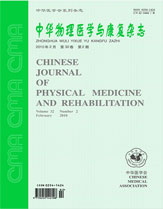
Chinese Journal of Physical Medicine and Rehabilitation

1979  to  Present  ISSN: 0254-1424

Articles

About

Save Email

Sort by

Best match
Relevance
PubYear
JournalTitle

DISPLAY OPTIONS

Format:

Per page:

Save citations to file

Selection:

Format:

Create file Cancel

Email citations

To:

Please check your email address first!

Selection:

Format:

Send email Cancel

2520

results

page

of 252

1

Cite

Cite

Copy

Share

Share

Copy

Effects of low frequency repetitive transcranial magnetic stimulation on motor function and excitability of motor cortex in patients with Parkinson's diseaseWANG Ming-wei, SUN Xiu-qiao, GU Ping, MA Xiao-wei,

Mingwei WANG ; Xiuqiao SUN ; Ping GU ; Xiaowei MA ; Yanmin LI ; Jihong GUO ; Haimin SUN ; Jun SHI

Chinese Journal of Physical Medicine and Rehabilitation.2009;31(8):529-533. doi:10.3760/cma.j.issn.0254-1424.2009.08.007

Objective To investigate the effects of low frequency repetitive transcranial magnetic stimulation (rTMS) on motor function and excitability of motor cortex in Parkinson's disease (PD) patients and to study the mechanism of PD from the electrophysiology. Methods Twenty-eight patients with PD received 1 Hz rTMS therapy for 15 d. Thirty normal volunteers were enrolled as controls. Unified Parkinson's Disease Rating Scale (UPDRS) and motor evoked potential (MEP) were adopted as assessment indicators. The excitability of motor cortex was assessed by rest motor threshold (RMT), central motor conduction time (CMCT) and the amplitude of MEP. Results The initial RMTs and CMCTs of PD patients were significantly lower than those of the controls, but MEP amplitudes were not significantly different. After rTMS treatment, motor function of PD patients improved, RMTs increased and CMCTs prolonged. Conclusion In PD patients, motor function disorder and increased motor cortical excitability were observed. Low frequency rTMS may inhibit these changes to some extent.

2

Cite

Cite

Copy

Share

Share

Copy

The effects of magnetothcrapy on local lymphocyte infiltration and neurocytes apoptosis after traumatic brain injury of rats

Yuping WANG ; Xiubao SONG ; Qisheng HONG ; Wei LI

Chinese Journal of Physical Medicine and Rehabilitation.2009;31(8):524-527. doi:10.3760/cma.j.issn.0254-1424.2009.08.005

Objective To study the neuro-protective effects of magnetotherapy after traumatic brain injury of rats. Methods Traumatic brain injury models were created in 20 rats and divided randomly into magnetotherapy and non-magnetotherapy groups. The rats in magnetotherapy group received magnetotherapy on the cranial surface of traumatized region twice daily, each for 20 min, over 10 d. The intensity of magnetic field was O. 15 T at the surface,and 0.1 T during spinning at 3000 rpm. The contraction strength of gastrocnemius of each rat's left hind limb was measured on the 11 th d after trauma. The rats were then sacrificed and brain tissue samples were taken. The infiltra-tions of CD4 + and CD8 + cells and apnptosis in the area around the injury lesion were observed with immunohisto-chemical methods. The relationship among the indexes was analyzed. Results At the 11th d after brain injury, the quantities of CD4 + and CD8 + cells and apoptotic cells in magnetotherapy group were all obviously less than those in non-magnetotherapy group. Average contraction strength of gastrocnemius in magnetotherapy group was significantly stronger than that in non-magnetotherapy group. Conclusions Magnetotherapy can relieve the secondary effects of brain injury and induce neuro-protection effects. This may involve decreasing the local infiltrations of CD4 + and CD8 + and apoptotic cells in the area around the injury lesion.

3

Cite

Cite

Copy

Share

Share

Copy

The effects of exercises on the expression of matrix metalloproteinase-9 in perihematomal brain tissue after intra cerebral hemorrhage in rats

Duozi WANG ; Fuqiang GUO ; Hongyan ZHANG ; Hao SUN ; Xiangrong SUN ; Xianrong ZENG ; Jinyu WANG ; Wenbing WU ; Fuqiong PAN

Chinese Journal of Physical Medicine and Rehabilitation.2009;31(8):509-513. doi:10.3760/ema.j.issn.0254-1424.2009.08.002

Objective To study the effects of exercises training on the recovery of neurological function and the expression of matrix metalloproteinase-9 (MMP-9) in perihematomal brain tissue after intra cerebral hemorrhage (ICH) in rats. Methods Sixty-four male adult Sprague-Dawley rats were randomly divided into exercises group and control group. ICH model was induced by autobiood injection. The rats in exercises group were given balance, grasping and walking exercises every day. The rats in control group exercised freely in standard cages. Neurological function in both groups was measured at the 3rd, 7th, 14th and 21st d. All the rats were sacrificed and the concentration of MMP-9 was measured with immunohistochemical method and in situhybridization. Results In both groups neurological function scores was lowest at the 3rd d, were highest at the 21st d. There was no significant difference between two groups at the 3rd d, but at the 7th, 14th and 21st d the differences between two groups were significant ( P < 0.05 ). In exercises group, MMP-9 expressed weakly at the 3rd d, and peaked at the 7th d, then declined gradually. In control group, MMP-9 expression peaked at the 3rd d, then decreased slowly. MMP-9 protein expression was in accord with mRNA. MMP-9 expression in exer-cises group was significantly different from control group on the 3rd and 7th d (P < 0.05 ) , but not at the 14th and 21st d ( P > 0.05 ). Conclusions Early exercises can inhibit the expression of MMP-9 that could play a role in protecting neurons. Daily exercises can stimulates the expression of MMP-9 so as to have a positive role in midstage of disease. MMP-9 may be involved in tissue remodelling and vascular repairing, which prompt neu-rofunction recovery.

4

Cite

Cite

Copy

Share

Share

Copy

The effects of respiratory training on pulmonary function in patients with chronic obstructive pulmonary disease

Zhanxiang LIU ; Weihua ZHANG ; Yanli LI ; Anmeng WEI ; Xiuzheng CAO

Chinese Journal of Physical Medicine and Rehabilitation.2009;31(7):474-476. doi:10.3760/cma.j.issn.0254-1424.2009.07.014

Objective To observe the effects of respiratory training on pulmonary function in the patients with chronic obstructive pulmonary disease(COPD). Methods A total of 74 patients with COPD were recruited and randomly divided into a treatment group and a control group with 37 subjects in each.The treatment group re-ceived respiratory training including lip-contracting breathing,abdominal breathing and breathing exercises for 6 months in addition to routine medical treatment.The control group received only routine medical treatment.Pulmona-ry function was evaluated before and after treatment. Results After treatment,both groups improved in terms of all the pulmonary function measures,showing increased vital capacity(VC),forced expiratory volume after 1 s(FEVl),peak forced expiratory flow rate(PEFR),as well as forced expiratory flow at 25%of Vcmax(V25),de-creased residual volume and residual volume as a percentage of total lung capacity(RV/TCL).The difference be-tween the two groups was significant with regard to the extent of improvement. Conclusion Respiratory training can improve the pulmonary function of COPD patients.

5

Cite

Cite

Copy

Share

Share

Copy

The clinical value of a triple stimulation technique in the diagnosis of benign monomelic amyotrophy

Ying XIAO ; Bingdi XIE ; Hui ZHAI

Chinese Journal of Physical Medicine and Rehabilitation.2009;31(7):463-465. doi:10.3760/cma.j.issn.0254-1424.2009.07.010

Objective To assess the dlagnostic value of a triple stimulation technique(TST)for benign monomelic amyotrophy(BMA). Methods A total of 15 BMA patients and 15 healthy control subiects were exam-ined with the TST.The latent periods,amplitudes,areas and durations of the negative waves of the compound muscle action potential(CMAP)and motor evoked potential(MEP)induced by the stimulation were recorded,and the de-creasing ratio of amplitudes and areas in the two groups were analyzed. Results There were significant differences in amplitude and area ratios between the BMA patients and the healthy control subjects.Both ratios were significantly lower in the BMA patients. Conclusion TST has some value in diagnosis of BMA.

6

Cite

Cite

Copy

Share

Share

Copy

Analysis of abnormal motor evoked potentials of the obese subjects induced by magnetic stimulation at T12 to L1

Shaoping WANG ; Zengfu LI ; Peichao TIAN ; Chengwen LI

Chinese Journal of Physical Medicine and Rehabilitation.2009;31(7):460-462. doi:10.3760/cma.j.issn.0254-1424.2009.07.009

Objective To study the abnormalities of motor evoked potentials(MEPs)obtained from obese subjects induced by magnetic stimulation at T12 to L1. Methods A total of 90 healthy subjects without any neuro-muscular system abnormalities were divided into 3 groups according to the body mass index(BMI).The control group consisted of 30 normal somatotype males(BMI<23),while the obese somatotype group(BMI>30)and weight-reduc-ing group(BMI>30)consisted of 30 obese somatotype males,respectively.The subjects in the control group and obese somatotype group maintain their usual lifestyle as before,while those in the weight-reducing group were reques-ted to lose weight by all means.MEPs were induced by using magnetic stimulation of 70%and 90%the output of the stimulator,respectively,at T12 to L1 and recorded from all the anbjcots at the time of admission to the study and one year later. Results At admission,the amplitude and conduction time of MEPs in obese somatotype group and weight-reducing group were significantly different from those in control group(P<0.05),when the magnetic stimula- tion of 70%the stimulator output was employed.but all the indexes were not difierent when 90%the stimulator uot- put were adopted(P>0.05).One year later,the amplitude and conduction time of MEPs in obese somatotype group were significantly different from those in control group and weight-reducing group(P<0.05),but all the indexes were not different within 3 groups when 90%the stimulator output was adopted(P>0.05). Conclusion Supra-maximal magnetic stimulation should be used for recording the MEPs from the obese subjects,or false positive result would be obtained.

7

Cite

Cite

Copy

Share

Share

Copy

Lower extremity muscle coordination in stroke patients revealed by surface electromyography

Zulin DOU ; Hongmei WEN ; Li JIANG ; Lei ZHU

Chinese Journal of Physical Medicine and Rehabilitation.2009;31(7):448-451. doi:10.3760/cma.j.issn.0254-1424.2009.07.006

Objective To explore the changes in muscle activity patterns in the lower leg during ankle dorsiflexion and plantarflexion in stroke patients. Methods Ten recovering stroke patients with hemiparesis(Pa- tient group)and ten age-and sex-matched healthy volunteers(Healthy control group)were studied.The subjects performed ankle dorsiflexion and plantarflexion synchronized with a visual cue while supine.Surface electrodes were applied over the anterior tibialis(TA),caput laterale musculi gastrocnemius(IGM),caput mediale musculi gas-troenemius(mGM)and soleus muscles(SOL)for integrated electromyography(iEMG).Results During ankle dorsiflexion,the TA,lGM,mGM and SOL of the affected side showed a significantly lower iEMG signal than the unaffected side.Activity of the lGM,mGM and SOL of the affected side were significantly lower than in the healthy controls.During ankle plantaflexion,the TA,lGM and mGM on the affected side had significantly lower activity than those of the healthy controls.The mGM of the affected side showed significantly lower muscle activity than the unaffeeted side.The SOL contraction ratio on the affected side was significantly higher than on the unaffected side and in the healthy control group. Conclusion Except for the soleus,there is an obvious decline in muscle activi-ty in the affected lower extremities of stoke patients during ankle dorsiflexion and plantaflexion.The soleus on the affected side becomes the primary plantarflexor in patients with stroke,rather than the caput laterale musculi gas-trocnemius or caput mediale musculi gastroenemius as in healthy subjects.

8

Cite

Cite

Copy

Share

Share

Copy

A preliminary study of type I collagen expression of bone marrow mesenchymal stem cells promoted by pulsed electromagnetic fields

Yong YANG ; Hua WU ; Chaoxiong TAO ; Feng LI ; Rui LI ; Wenchun ZHAO

Chinese Journal of Physical Medicine and Rehabilitation.2009;31(7):442-445. doi:10.3760/cma.j.issn.0254-1424.2009.07.004

Objective To study the influence of pulsed electromagnetic fields on the expression of type I collagen by bone marrow mesenchymal stem cells and it's mechanism. Methods The bone marrow mesenchymal stem cells of SD rats were isolated and cultured in vitro.The third passage cells were harvested and exposed to pulsed electromagnetic fields(PEMFs)at 15 Hz and 1 mT 8 h/d for 3 days.A semi-quantitative RT-PCR technique was used to measure the type I collagen mRNA expression;ELISA and immunohistochemitistry techniques were used to measure type I collagen expression.Inhibitors and promoters of the cAMP-dependent protein kinase A(cAMP-PKA)pathway were added.After the cAMP-PKA pathway had been inhibited or promoted,the effects of the PEMF on type I collagen expression were measured again using ELISA and immunohistoehemistry.Results PEMFs at 15 Hz and 1 mT induced significant promotion of the expression of type I collagen(P≤0.01)in comparison with the controls. The type I collagen expression was reduced when the cAMP-PKA pathway inhibitor H-89 was added,and raised when the promoter 8-Br-cAMP was added.Conclusion PEMFs at 15 Hz 1 mT can promote type I collagen expression of bone marrow mesenchymal stem cells.and the effect is correlated with the cAMP-PKA pathway.

9

Cite

Cite

Copy

Share

Share

Copy

The effects of rehabilitation on learning,memory and expression of NCAM in rats with vascular dementia

Zhenyong FAN ; Lina CHEN ; Linfeng XU ; Ya ZONG ; Jianyong HU ; Xianghua YU

Chinese Journal of Physical Medicine and Rehabilitation.2009;31(6):361-365. doi:10.3760/cma.j.issn.0254-1424.2009.06.001

Objective To investigate the effect of rehabilitation training on learning and memory ability and the expression of neural cell adhesion molecule(NCAM)in rats modeling vascular dementia.Methods Foay-five female Sprague-Dawley rats were randomly assigned to a rehabilitation group(20 rats),an immobilization group(20 rats),or a sham-operation group(5 rats).The experimental vascular dementia model was established by repeatedly clipping the common carotid artery to induce repetitive isehemia-reperfusion,and by reducing blood pressure with intra-abdominal injection of sodium nitroprusside.The rats' learning and memory were tested on the 27th and 28th days after the operation using a water-maze step-down avoidance test.A RT-PCR technique was used to detect NCAM expression around the hippocampal area at different times after the operation.Results The rehabilitation group rats showed significantly better learning and memory ability than those in the immobilization group.NCAM was also more strongly expressed in their hippocampi than in those of the immobilization group and sham-operation group.Conclusion Rehabilitation can accelerate recovery of learning and memory ability in rats,and the mechanism possibly is related to the increase of NCAM expression in the hippocampus.

10

Cite

Cite

Copy

Share

Share

Copy

The prevention and cure of postoperative neck axial symptoms after open-door laminoplasty for cervical spondylotic myelopathy

Junming CAO ; Dalong YANG ; Yong SHEN ; Wenyuan DING ; Liu YANG ; Jiaxin XU

Chinese Journal of Physical Medicine and Rehabilitation.2009;31(7):468-471. doi:10.3760/cma.j.issn.0254-1424.2009.07.012

Objective To evaluate the effectiveness of rehabilitative treatment after extended open-doorlaminoplasty of the cervical spine for cervical spondylotic myelopathy.Methods Sixty patients(mean age 61.7 ±12 years)who had undergone extended open-door laminoplasty of the cervical spine for cervical spondylotic myelopa-thy were studied for an average of 40.7 months.They are divided into a rehabilitation group and a control group with 30 patients in each.The patients in the rehabilitation group received systematic rehabilitation therapy pre-and post-operation.In both groups,the neurological recovery rate,the cross-sectional areas of the cervical posterior muscles,and the incidence of axial symptoms and post-operative complications were recorded and compared. Results The wounds of patients in the rehabilitation treatment group healed with no complicating infections,but the wounds of three patients in the control group became infected.There was no statistically significant difference in neurological recovery between the two groups.In the rehabilitation treatment group,the rate of atrophy and the cross-sectional areas of the cervical posterior muscles were significantly lower than in the control group.At the same time,the rate of neck axial symptoms was 23% in the rehabilitation group but 60%in the control group.a difference which was statistically significant. Conclusions Although rehabilitation treatment pre-and post-operation may not speed up neurological recovery,it may prevent postoperative atrophy of the cervical extensors and lower the incidence of neck axial symptoms.

Country

China

Publisher

中华医学会

ElectronicLinks

http://www.cjpmr.cn/ch/

Editor-in-chief

E-mail

cjpmr@tjh.tjmu.edu.cn

Abbreviation

Chinese Journal of Physical Medicine and Rehabilitation

Vernacular Journal Title

中华物理医学与康复杂志

ISSN

0254-1424

EISSN

Year Approved

2008

Current Indexing Status

Currently Indexed

Start Year

1979

Description

历史沿革【现用刊名:中华物理医学与康复杂志;曾用刊名:中华物理医学杂志;创刊时间:1979】,该刊被以下数据库收录【CA 化学文摘(美)(2009);CBST 科学技术文献速报(日)(2009);中国科学引文数据库(CSCD—2008)】,核心期刊【中文核心期刊(2008);中文核心期刊(2004);中文核心期刊(2000);中文核心期刊(1992)】。

Related Sites

WHO WPRO GIM

Help Accessibility
DCMS Web Policy
CJSS Privacy Policy

Powered by IMICAMS( 备案号: 11010502037788, 京ICP备10218182号-8)

Successfully copied to clipboard.