

Main content 1 Menu 2 Search 3 Footer 4
+A
A
-A
High contrast
HOME JOURNAL JOURNAL SELECTION NETWORK HELP ABOUT

Journal Selection Criteria and Standards

WPRIM Journal Selection Criteria (August 2026)

NJSC Philippines Selection Criteria (for Philippine-based journals only)

Minimum standards for the suspension and removal of WPRIM approved journals

Application and Indexing Process

Application and Submission Process for WPRIM Indexing

Journal Content Management

Candidate Journal Selection and Data Creation and Management System

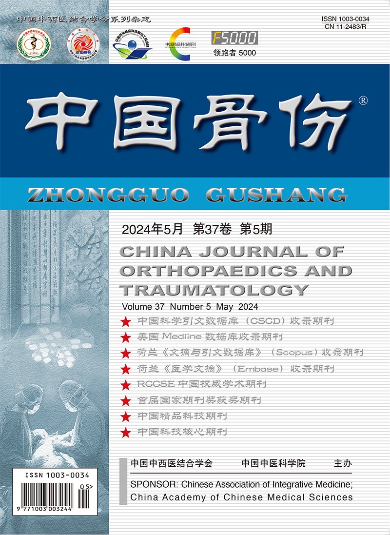
China Journal of Orthopaedics and Traumatology

1987  to  Present  ISSN: 1003-0034

Articles

About

Save Email

Sort by

Best match
Relevance
PubYear
JournalTitle

DISPLAY OPTIONS

Format:

Per page:

Save citations to file

Selection:

Format:

Create file Cancel

Email citations

To:

Please check your email address first!

Selection:

Format:

Send email Cancel

4786

results

page

of 479

1

Cite

Cite

Copy

Share

Share

Copy

Percutaneous kyphoplasty for the treatment of osteoporotic vertebral compression fracture with degenerative scoliosis.

Hang-bo QU ; Pei-jian TONG ; Wei-feng JI ; Ju LI

China Journal of Orthopaedics and Traumatology.2016;29(1):38-40.

OBJECTIVETo evaluate the therapeutic effects of percutaneous kyphoplasty (PKP) for the treatment of osteoporotic vertebral compression fracture (OVCF) with degenerative scoliosis.

METHODSFrom March 2009 to March 2014,121 patients undergoing PKP for the treatment of osteoporotic vertebral compression fracture (OVCF) with degenerative scoliosis were retrospective analyzed. There were 41 males and 80 females,ranging in age from 56 to 92 years with an average of 73.2 years. Preoperative and postoperative 3 d respectively pain visual analogue scale (visual analogue score, VAS) and Oswestry Disability Index assessed pain and functional recovery of patients and preoperative and postoperative 3 d lumbar lordosis, scoliosis Cobb angle were analyzed.

RESULTSAll the patients were followed up, and the duration ranged from 6 to 24 months, with a mean time of 11 months. All the patients achieved success in operation without serious complications. The operation time ranged from 30 to 65 min with an average of 42.2 min. Local leakage of bone cement was not found in canal. The VAS and Oswestry Disability Index at the 3rd day after operation decreased significantly compared with those of the preoperative (P < 0.05), and the lumbar lordosis and scoliosis Cobb angle also improved significantly compared with those of the preoperative (P < 0.05).

CONCLUSIONPercutaneous kyphoplasty (PKP) for the treatment of thoracolumbar scoliosis osteoporotic fracture can significantly improve patients spinal deformity, pain relief, which is worth of recommending.


Aged ; Aged, 80 and over ; Female ; Fractures, Compression ; surgery ; Humans ; Kyphoplasty ; methods ; Male ; Middle Aged ; Osteoporotic Fractures ; surgery ; Scoliosis ; surgery ; Spinal Fractures ; surgery ; Visual Analog Scale
Aged ; Aged, 80 and over ; Female ; Fractures, Compression ; surgery ; Humans ; Kyphoplasty ; methods ; Male ; Middle Aged ; Osteoporotic Fractures ; surgery ; Scoliosis ; surgery ; Spinal Fractures ; surgery ; Visual Analog Scale

2

Cite

Cite

Copy

Share

Share

Copy

Case-control studies of two kinds of method for the treatment of lumbar tuberculosis with psoas abscess.

Qi WANG ; Ming HU ; Yuan-zheng MA ; Xiao-bo LUO

China Journal of Orthopaedics and Traumatology.2016;29(1):33-37.

OBJECTIVETo compare two kinds of method for treating lumbar tuberculosis with psoas abscess, to provide reference for clinical reasonable select of therapy treatment.

METHODSFrom January 2010 to January 2013,42 patients with lumbar tuberculosis combined with psoas abscess with obvious surgical indications were enrolled, including 24 males and 18 females with an average age of (38.5 ± 10.2) years old ranging from 21 to 63 years old. All patients were followed up for 18 to 24 months with an average of 20.9 months. Twenty-two patients underwent posterior vertebral body lesions cleared, bone graft fusion and internal fixation and percutaneous puncture catheter drainage for treatment of psoas major abscess as group A, and twenty patients underwent one-stage extraperitoneal approach to remove abscess, posterior vertebral body lesions cleared, bone graft fusion and internal fixation as group B. The operative time, loss of blood, length of hospital stay, clinical cure rate and other clinical results for the two groups were analyzed and compared.

RESULTSThe loss of blood was (452.3 ± 137.6) ml in group A and (603.5 ± 99.6) ml in group B, there was significant statistical difference (P < 0.05). The time of operation was (193.6 ± 91.2) min in group A and (230.5 ± 56.6) min in group B, there was significant statistical difference (P < 0.05). The time of operation and the loss of blood in group A were obviously less than which in group B. In group A 20 cases were cured and 2 cases relapsed, 19 cases were cured and 1 case relapsed in group B, there was no significant statistical differences between two groups regarding cure rate with chi-square test (χ² = 0.000, P = 1.000). All patients in two groups obtained good clinical curative effect. There were no significant statistical difference between two groups regarding for length of hospital stay with t-test (P > 0.05).

CONCLUSIONLumbar spinal tuberculosis with psoas abscess is not absolute indications for anterior open operation. Compared with the combined anterior and posterior surgical procedure, the percutaneous puncture catheter drainage combined with posterior debridement, interbody fusion and internal fixation can achieve the same clinical effect but less trauma for the patients.


Adult ; Case-Control Studies ; Debridement ; Female ; Humans ; Lumbar Vertebrae ; surgery ; Male ; Middle Aged ; Psoas Abscess ; etiology ; surgery ; Spinal Fusion ; Tuberculosis, Spinal ; complications ; surgery ; Young Adult

Adult ; Case-Control Studies ; Debridement ; Female ; Humans ; Lumbar Vertebrae ; surgery ; Male ; Middle Aged ; Psoas Abscess ; etiology ; surgery ; Spinal Fusion ; Tuberculosis, Spinal ; complications ; surgery ; Young Adult

3

Cite

Cite

Copy

Share

Share

Copy

Clinical studies of pedicle screw-rod fixation of thoracolumbar burst fractures through posterior unilateral approach after vertebrae corpectomy fusion.

Yong-jun HUA ; Ren-yan WANG ; Zhi-hui GUO ; Cun-hong SHU ; Chao-hua LI

China Journal of Orthopaedics and Traumatology.2016;29(1):27-32.

OBJECTIVETo compare the clinical curative effect of thoracolumbar burst fracture treated by the posterior unilateral approach corpectomy fusion screw-rod fixation and anterior corpectomy bone fusion screw plate fixation.

METHODSFrom January 2008 to May 2014,36 cases of thoracolumbar burst fracture underwent operation of decompression, fusion, and internal fixation was retrospective analyzed. Among them, 16 patients were treated through posterior approach as posterior group, including 13 males and 3 females aged from 37 to 62 years old; 9 cases caused by falling injury, 3 cases by traffic accident injury,4 cases by heavy aboved;the injury segment was on T₁₂ in 2 cases, L₁ in 5 cases, L₂ in 7 cases, L₃ in 2 cases; according ASIA grade, 3 cases were grade A, 2 cases were grade B, 2 cases were grade C, 5 cases were grade D, 4 cases were grade E; the time between injury and operation ranged from 5 to 15 days. Other 20 patients were treated through anterior-lateral approach as anterior-lateral group, including 15 males and 5 females with age from 27 to 62 years old; 12 cases caused by falling injury, 4 cases by traffic accident injury, 4 cases by heavy aboved; the injury segment was on T₁₂ in 2 cases, L₁, in 7 cases, L₂ in 9 cases, L₃ in 2 cases; for ASIA grade: 4 cases were grade A, 2 cases were grade B, 4 cases were grade C, 6 cases were grade D, 4 cases were grade E; the time between injury and operation ranged from 4 to 12 days. The operation time, bleeding during operation and postoperative drainage volume were observed in two groups,and the changes of nerve function of ASIA grade, clinical efficacy,improved degree of thoracic and lumbar lordosis,and bony fusion were compared between two groups.

RESULTSAll patients were followed up from 12 to 24 months with an average of (15.8 ± 3.3) months. The operation time, bleeding during operation, and postoperative drainage volume had no significant different between two groups (P > 0.05). As compared with preoperative, ASIA grade of two groups at last follow-up had statistically significantly different (P < 0.01), the neural function of two groups after operation was recovered for different extent. The JOA score of two groups was compared between last follow-up and preoperative, the difference had statistically significant (P < 0.01), the two groups showed good clinical effect. The clinical results of ASIA grade, JOA score and RIS had no significant differences between two groups. All patients of two groups were obtained fusion. Thoracic and lumbar lordosis angle improvement degree had no significant difference between two groups ,it bad significant difference had statistical significance compared with preoperative, the two approaches could effectively restore the spinal sequence.

CONCLUSIONFor patients with thoracolumbar burst fracture just treated by anterior decompression and reconstruction of anterior column, according to the degree of operation performer' skill proficiency and the patient' condition to choose, but for patients must performed the spinal canal decompression anterior and posterior, the three column-reconstruction to required anterior-posterior approach, the posterior unilateral approach corpectomy fusion screw-rod fixation obviously shorten operation time, reduce the operation wound, it is worth the clinical promotion.


Adult ; Decompression, Surgical ; methods ; Female ; Humans ; Lumbar Vertebrae ; injuries ; surgery ; Male ; Middle Aged ; Pedicle Screws ; Reconstructive Surgical Procedures ; Retrospective Studies ; Spinal Fractures ; surgery ; Spinal Fusion ; methods ; Thoracic Vertebrae ; injuries ; surgery

Adult ; Decompression, Surgical ; methods ; Female ; Humans ; Lumbar Vertebrae ; injuries ; surgery ; Male ; Middle Aged ; Pedicle Screws ; Reconstructive Surgical Procedures ; Retrospective Studies ; Spinal Fractures ; surgery ; Spinal Fusion ; methods ; Thoracic Vertebrae ; injuries ; surgery

4

Cite

Cite

Copy

Share

Share

Copy

Comparison between volar and radial column approach by plate fixation for the treatment of unstable fracture of distal radius: a Meta-analysis.

Ye-cheng LI ; Wei ZHANG ; Shou-zheng LIU ; Cheng-liang ZHANG ; Hong-jun WANG

China Journal of Orthopaedics and Traumatology.2016;29(1):21-26.

OBJECTIVETo assess the clinical effectiveness of volar and radial column approach by plate fixation for the treat- ment of unstable fracture of distal radius.

METHODSAccording to Cochrane Systematic Review, Medline, Embase, Cochrane Li- brary, CNKI and CBM, randomised controlled trials (RCTs) of volar and radial column approach by plate fixation for the treat- ment of unstable fracture of distal radius were searched for from 1966 to 2014. Data analysis was performed with the Cochrane Collaboration's RevMan 5.0 software.

RESULTSTotally 391 patients of 6 RCTs and 2 retrospective cohort studys were included and divided into volar plate group (187 cases) and radial column plate group (204 cases). Meta-analysis result showed: compared with radial column plate group, volar plate group had significant difference in recovery of wrist function [SMD = 0.74, 95% CI (0.47, 1.01), P < 0.00001], Gartland-Werley scores [SMD = -1.39, 95% CI (-2.24, -0.53), P = 0.001], postoperative neural in- jury [OR = 3.67, 95% CI (1.37, 9.84), P = 0.01 1 and postoperative wrist pain [OR = 0.32, 95% CI (0.13, 0.74), P = 0.008]. But no significant difference was identified in DASH scores [SMD = -0.36, 95% CI (-0.97, 0.26), P = 0.25], radiographic result assess- ment [SMD = -0.18, 95% CI (-0.53, 0.16), P = 0.3], postoperative grip strength [SMD = 0.71, 95% CI (-0.12, 1.54),P = 0.09], postoperative tendinous damage [OR = 0.31, 95% CI (0.10, 0.98), P = 0.05] and carpal tunnel syndrome [OR = 0.96, 95% CI (0.63, 1.48), P = 0.87].

CONCLUSIONCompared with radial column plate internal fixation, volar approach plate fixation for treat- ment of distal radius intra-articular fracture has advantage of recovery of joint functionand. However, the volar approach plate fix- ation was associated with a higher risk of long-term complications than the radial column approach plate fixation.


Bone Plates ; Fracture Fixation, Internal ; methods ; Humans ; Radius Fractures ; surgery

Bone Plates ; Fracture Fixation, Internal ; methods ; Humans ; Radius Fractures ; surgery

5

Cite

Cite

Copy

Share

Share

Copy

Treatment of Colles fracture with manipulative closed reduction and U-shaped gypsum functional fixation.

Su JIAN-CAI

China Journal of Orthopaedics and Traumatology.2016;29(1):18-20.

OBJECTIVETo investigate the effect of U-shaped gypsum functional fixation after closed manipulative reduction for treatment of Colles' fractures.

METHODSFrom January 2011 to April 2014,47 cases of Colles fracture were treated by closed reduction and U-shaped gypsum functional fixation including 12 males and 35 females with an average age of 54.8 years old ranging from 8 to 72 years old. The time from injury to treatment was 40 min to 3 d. The patients were closed fractures without neurovascular injury. After manual reduction and U-shaped gypsum functional fixation, the thumb of injury hand were traction by contralateral hand, and other fingers of injury hand were taken flexion to exercise grip function. According to the situation of fracture healing, plaster was removed at 4 to 8 weeks' fixation, the wrist joint functional exercise was strengthened after plaster removed.

RESULTSAll patients were followed up from 6 to 12 months with an average of 6.4 months, the fracture healing time was 4 to 8 weeks. According to the Cooney wrist function scoring: the result was excellent in 44 cases, good in 2 cases, 1 case.

CONCLUSIONU-shape gypsum functional fixation for treatment of Colles fracture limits the activity of thumb and extensor tendon, can reduce shortening of radial by traction of the injury thumb with the contralateral hand; it is good for blood circulation of hand and wrist, and swelling.


Adolescent ; Adult ; Aged ; Child ; Colles' Fracture ; therapy ; Female ; Fracture Fixation ; methods ; Humans ; Male ; Manipulation, Orthopedic ; methods ; Middle Aged

Adolescent ; Adult ; Aged ; Child ; Colles' Fracture ; therapy ; Female ; Fracture Fixation ; methods ; Humans ; Male ; Manipulation, Orthopedic ; methods ; Middle Aged

6

Cite

Cite

Copy

Share

Share

Copy

Quantitative evaluation of Colles' fracture by Multislice CT multiplanner reconstruction: a feasibility study.

Jiu-zun CHEN ; Feng LIN ; Sheng-fa ZHOU ; Wei CHEN ; Meng-nan WAN ; Jia-wei HE ; Xian-qiu ZHENG ; Min DAI

China Journal of Orthopaedics and Traumatology.2016;29(1):13-17.

OBJECTIVETo investigate the feasibility and reliability on the quantitative evaluation of Colles' fracture by multislice CT (MSCT) multiplanner reconstruction (MPR).

METHODSA total of 36 patients with Colles' fracture from July 2011 to July 2014 were investigated in this study. There were 11 males and 25 females with a mean age of (42.5 ± 5.4) years old (ranged 35 to 72 years). All the patients underwent anteroposterior and lateral X-ray films and MSCT scans on wrist joints within 2 days after trauma. Images were sent to the workstation through picture archiving and conserving system (PACS). One associate chief physician independently and respectively measured the dorsal intercalation depth of distal fracture block, palmar angle and dislocation degree of wrist articular surface collapse on anteroposterior and lateral X-ray film and MSCT-MPR. The time interval between the two measurements was 2 weeks. All the data between the first and second measurement on X-ray and MPR and the mean value between the X-ray and MPR was examined with paired t-test. The pearson analyzed their correlation.

RESULTSAmong the 35 cases, 35 cases of palmar angle, 21 cases of intercalation depth and 16 cases of dislocation of wrist articular surface collapse could be measured on both X-ray and MPR. For the above parameters, the first measurement results were (12.5 ± 3.6)°, (4.5 ± 2.1) mm, (3.7 ± 1.6) mm and the second measurement results were (4.8 ± 2.2)°, (6.4 ± 3.6) mm, (2.5 ± 1.2) mm on X-ray films respectively. The first measurement results on MPR were (14.5 ± 5.3)°, (4.2 ± 1.2) mm, (5.7 ± 2.3) mm, and the results were (13.2 ± 2.6)°, (4.7 ± 2.2) mm, (4.6 ± 2.1) mm for the second measurement respectively. The three parameters between the first and second measurement on plain film had statistical difference and low correlation (r = 0.681, 0.640, 0.345, P < 0.05). The data between the first and second measurement on MPR showed that the dislocation degree of wrist articular surface collapse had statistical difference (P < 0.05) and no statistical significance was found for the other two parameters (P > 0.05), with the moderate correlation (r = 0.954, 0.854, 0.642). The three parameters had low or moderate correlation with each other on X-ray (r = 0.454, 0.532, 0.378, P < 0.05), compared with the mean value on MPR.

CONCLUSIONUsing MSCT MPR images may carry on the multiple parameter measurement of Colles fracture, to make quantitative evaluation, and repeated measurement is better reliability.


Adult ; Aged ; Colles' Fracture ; diagnostic imaging ; Feasibility Studies ; Female ; Humans ; Image Processing, Computer-Assisted ; Male ; Middle Aged ; Multidetector Computed Tomography ; methods

Adult ; Aged ; Colles' Fracture ; diagnostic imaging ; Feasibility Studies ; Female ; Humans ; Image Processing, Computer-Assisted ; Male ; Middle Aged ; Multidetector Computed Tomography ; methods

7

Cite

Cite

Copy

Share

Share

Copy

Clinical research of percutaneous K-wires fixation after manipulative reduction combining with gypsum or splint fixation for treatment of the Barton fractures of aged people.

Jian-liang CHEN ; Long-jun ZHANG ; Yong XU ; Shao-bing ZHU ; Xiao-dong ZHENG

China Journal of Orthopaedics and Traumatology.2016;29(1):8-12.

OBJECTIVETo investigate the feasibility of using percutaneous K-wires fixation after manipulative reduction combining with gypsum or splint fixation for treatment of the Barton fracture of aged people.

METHODSFrom June 2011 to May 2013, 91 patients with closed and fresh Barton fracture were analyzed retrospectively which information was complete. According to different methods these patients were divided into three groups: 33 patients in group A were treated by simplex fixation of gypsum or splint after manipulative reduction, including 20 males and 13 females with an average age of (63.27 ± 5.83) years old; 35 patients in group B were treated by percutaneous K-wires fixation after manipulative reduction combining with gypsum or splint fixation, including 23 males and 12 females with an average age of (64.25 ± 4.53) years old; 23 patients in group C were treated by open reduction and internal fixation including 15 males and 8 females with an average age of (64.04 ± 4.83) years old.

RESULTSAll the patients were followed up for 10 to 16 months with an average of 12 months by the method of recheckging in outpatient clinic. According Dienst scoring system: in group A, the result was excellent in 15 cases, good in 7 cases, fair in 11 cases; in group B, excellent in 25 cases, good in 6 cases, fair in 4 cases; in group C, excellent in 16 cases, good in 5 cases, fair in 2 cases. The excellent and good rate of group C was better than that of group B and A, further more group B is better than group A. According to X- ray standard, in group A, 26 patients were acceptable, 7 patients were unacceptable; in group B,28 patients were acceptable, 7 patients were uacceptable; in group C, 19 patients were acceptable,4 patients were unacceptable; there were no statistical significant difference (P > 0.05). There were 18 patients with emerging recrispation in group A, 10 patients in group B, 6 patients in group C, the result of group B and C was better than that of group A, and there was no statistically significant between group B and C (P > 0.05).

CONCLUSIONUsing percutaneous K-wires fixation after manipulative reduction combining with gypsum or splint fixation for treatment of the Barton fracture of aged people is an effective method, it has advantages of micro-invasived, reliable fixation, less complication, not need to fix wrist joint and early functional exercise.


Aged ; Bone Wires ; Female ; Humans ; Male ; Manipulation, Orthopedic ; methods ; Middle Aged ; Radius Fractures ; therapy ; Retrospective Studies ; Splints

Aged ; Bone Wires ; Female ; Humans ; Male ; Manipulation, Orthopedic ; methods ; Middle Aged ; Radius Fractures ; therapy ; Retrospective Studies ; Splints

8

Cite

Cite

Copy

Share

Share

Copy

Predictive value of radiological parameter for re-displacement of conservative treatment of distal radius fracture.

Fei-ya ZHOU ; Wei-yang GAO ; Liang-fu JIANG ; Yong-huan SONG ; Zheng-lin CHI ; Jing-quan YANG ; Jian DING ; Ting-gang CHU

China Journal of Orthopaedics and Traumatology.2016;29(1):4-7.

OBJECTIVETo analyze the radiological parameters of the conservatively in treating distal radius fracture and investigate whether the final re-displacement can be predicted after primary reduction.

METHODSFrom January 2013 to June 2014,212 patients with distal radial fracture conservatively were treated in our hospital, 107 patients of them were excluded because of their incomplete radiological parameters;the remaining 105 patients were available for radiological were assessed after injury, there were 56 male patients and 49 female patients in this study, the average age of the patients was 51 years old (ranged from 22 to 80 years). According to AO classification, there were 47 cases of type A2 and C1, and 58 cases of type A3, C2, C3. All patients were treated by closed reduction and below-elbow cast immobilization for 4 to 6 weeks. All patients were followed up for 3 to 6 months (means 4.5 months) by X-ray, all fractures were healed. Standard AP and lateral radiographic examination was conducted before reduction and after reduction and bony consolidation,the dorsal angulation and the radial angle were measured at each time point. The linear regression was used for the analysis to find out whether the final re-displacement can be predicted after primary reduction.

RESULTSAmong 105 patients,the significant correlations were found for the dorsal angulation between the reduction time and the end time (r = 0.82) and for the radial angulation between the reduction time and end time (r = 0.85).

CONCLUSIONThe dorsal angulation and the radial angulation after complete healing can be predicted from linear the regression functions. Due to the possibility of predicting the end result, whether the fracture should receive further conservative treatment or surgical treatment can be decided immediately.


Adult ; Aged ; Aged, 80 and over ; Female ; Humans ; Male ; Middle Aged ; Predictive Value of Tests ; Radiography ; Radius Fractures ; diagnostic imaging ; therapy

Adult ; Aged ; Aged, 80 and over ; Female ; Humans ; Male ; Middle Aged ; Predictive Value of Tests ; Radiography ; Radius Fractures ; diagnostic imaging ; therapy

9

Cite

Cite

Copy

Share

Share

Copy

Thinking of Chinese and western medicine treatment for distal radius fractures.

Yong ZHAO

China Journal of Orthopaedics and Traumatology.2016;29(1):1-3.


Fracture Fixation ; Fracture Healing ; Humans ; Medicine, Chinese Traditional ; Radius Fractures ; therapy

Fracture Fixation ; Fracture Healing ; Humans ; Medicine, Chinese Traditional ; Radius Fractures ; therapy

10

Cite

Cite

Copy

Share

Share

Copy

Bottleneck and development trend of bone xenograft for the treatment of bone defect.

Bao-liang LI ; Lei LIU ; Wen-bo ZHAO ; Fu-jun LUAN ; Qin LI

China Journal of Orthopaedics and Traumatology.2015;28(12):1166-1170.

Bone xenograft bone for the treatment of bone defect is one of the current research focus, which has advantages of extensive sources, low cost, simple preparation method. While the process of single bone xenograft bone in repairing bone defect is very long, and the clinical outcome is not satisfactory. The main problems focus on formation of bone and vascularization. Reconstituted bone xenograft combined with cells and xenogenic bone material could promote vascularization and bone fusion in vivo, thus achieve a clinical effect of autogenous bone in repairing bone defect.
Bone Transplantation ; methods ; Bone and Bones ; blood supply ; Humans ; Transplantation, Heterologous

Bone Transplantation ; methods ; Bone and Bones ; blood supply ; Humans ; Transplantation, Heterologous

Country

China

Publisher

中国中西医结合学会;中国中医研究院

ElectronicLinks

http://www.zggszz.com

Editor-in-chief

E-mail

zggszz@sina.com

Abbreviation

China Journal of Orthopaedics and Traumatology

Vernacular Journal Title

中国骨伤

ISSN

1003-0034

EISSN

Year Approved

2008

Current Indexing Status

Currently Indexed

Start Year

1987

Description

历史沿革【现用刊名:中国骨伤;创刊时间:1987】,该刊被以下数据库收录【CA 化学文摘(美)(2009);Pж(AJ) 文摘杂志(俄)(2009)】,期刊荣誉【中科双奖期刊】。

Related Sites

WHO WPRO GIM

Help Accessibility
DCMS Web Policy
CJSS Privacy Policy

Powered by IMICAMS( 备案号: 11010502037788, 京ICP备10218182号-8)

Successfully copied to clipboard.